|

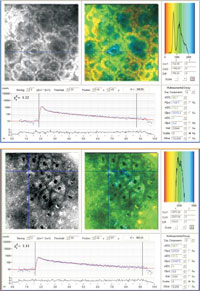
Fluorescence intensity image, false-color coded FLIM image, fluorescence lifetime histogram and fluorescence decay kinetics at a particular position
|
|
Besides 3D fluorescence imaging by optical sectioning and monitoring of the fluorescence intensity, DermaInspect® can also provide measurements on the lifetime of the autofluorescence with high submicron spatial as well as 250 ps temporal resolution giving the images an additional 4th dimension. Using time correlated single photon counting (TCSPC) DermaInspect® can perform fluorescence lifetime imaging (FLIM) in different tissue depths. The further parameter provides information on the type of the fluorescent biomolecule and its interaction with the microenvironment. For example, typical fluorescence lifetimes of free NADH, NADH-Protein complexes and porphyrin monomers are 200 ps, 2 ns, and 10 ns, respectively.
|
