| Applications |
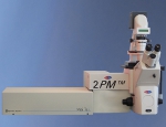
| The 2PM TM is a High Tech tool for three-dimensional life cell microscopy. Applications include the detection of fluorescent proteins in transfected cells and transgenic small animals. Furthermore, label free imaging can be performed based on two-photon excitation of endogenous fluorophores such as NADPH, flavoproteins, melanin, porphyrins, keratin, elastin, and collagen. |
 |
| (left, middle) optical sections through the cornea, autofluorescence (red), SHG (blue/collagen); (right) cancer cells |
| The microscope can be expanded to a 4D and 5D imaging tool, respectively, by adding Fluorescence Lifetime and spectral information. FLIM enables the mapping of fluorescence decay times in cells with submicron spatial and 50 ps / 250 ps temporal resolution even in tissue depths of 2 mm. This allows to study Förster (Fluorescence) Resonance Energy Transfer (FLIM-FRET) between fluorescent proteins as well as to distinguish different fluorophores and to probe the microenvironment. | |

